Monday 17 April 2017

DEEP VEIN THROMBOSIS


DEEP VEIN THROMBOSIS . 


DEFINITION  : Deep vein thrombosis occurs when blood clot forms and blocks a deep vein . Most commonly a vein of the leg or pelvis is affected . Every year , 1 out of 1000 person is afflicted among the populations of the Europe and the US . The risk increases with age . Women are affected more often than men . The most significant and dangerous complication of deep vein thrombosis is pulmonary embolism

COMPLICATION THROUGH PULMONARY EMBOLISM : When a clot detaches from the vesel wall , it may travel with the bloodstream into the pulmonary circulation and causes heart failure by blocking a pulmonary artery . Symptoms of pulmonary embolism includes shortness of breath , chest pain , coughing , restlessness, and paleness . 
DEVELOPMENT OF DISEASE : Damage to the vessel walls , deceleration of blood flow and a change in blood composition lead to an aggregation ( clumping together ) of the blood platelets ( thombocytes ) . The clot which have formed is enmeshed in a network of protein fibers ( fibrin ) , in which red blood cells may accumulate . 

Image result for damage of blood vessel


RISK FACTORS
:
The main factors contributing to the formation of deep vein thrombosis
are :

 - Restricted mobility through plaster casts ; paralyses and injuries
 - Lack of exercise due to restricted mobility  - Increased susceptibility to clotting after surgical interventions 
 - Pregnancy or chilbed  - Vein obstruction due to obesity or tumors 
 - Use of oral contraceptives , particularly in combination with nicotine consumption . 


FUNCTION OF MUSCLE PUMP :

 
A) Leg vein in lying position ; venous valves are open , the blood will flow freely to the heart.

B) Leg vein in standing position , muscle contraction phase : The venous valves above the contracted muscle fibers open . the blood flows towards the heart . The lower valve closes and prevents blood from flowing in the opposite direction ( retrograde flow ) . 

C) Leg vein in standing position , muscle relaxation phase : The venous valves in the area of relaxed muscle fibers opens , the blood flows towards the heart . The column of the blood closes the valve from above and prevents blood from flowing in opposite direction
( retrograde flow ) . 


Related image

SIGN & SYMPTOMS Pain or tenderness, swelling, warmth, redness or discoloration, and distention of surface veins, although about half of those with the condition have no symptoms. Signs and symptoms alone are not sufficiently sensitive or specific to make a diagnosis, but when considered in conjunction with known risk factors, can help determine the likelihood of Deep vein thrombosis . In most suspected cases, DVT is ruled out after evaluation, and symptoms are more often due to other causes, such as cellulitisBaker's cyst, musculoskeletal injury, or lymphedema. Other differential diagnoses include hematoma, tumors, venous or arterial aneurysms, and connective tissue disorders . 
PREVENTATION OF DVT : The main measure for preventing deep vein thrombosis is to support the venous bloodstream to the heart . This normally occurs through activation of the calf muscle pump while walking . For bedridden persons , the following measures help to prevent thrombosis : 

- Injections of anticoagulants
- Compressions stockings
- Physiotherapeutic mobility exercises in bed
- Earliest possible mobilization 

TREATMENT : Immobilization of the patient , administration of compression bandages , administration of anticoagulant drugs and painkillers are measures taken to prevent pulmonary embolism and the formation of new blood clots and to minimize swelling and 
pain . 


Image result for treatment of deep vein thrombosis in leg


PREVENTATION OF PULMONARY EMBOLISM : If pulmonary embolism occurs repeatedly despite administration of anticoagulants , a so called vena cava umbrella filter can be inserted into inferior vena cava through clavicular or femoral vein .This filter prevents clots from travelling from the leg or pelvic veins into the lungs . 


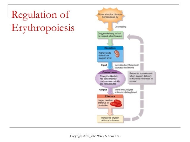
THE REGULATION OF ERYTHROPOIESIS

Image result for regulation of ERYTHROPOIESIS


Erythropoiesis  is the process which produces red blood cells (erythrocytes). It is stimulated by decreased oxygen in circulation, which is detected by the kidneys, which then secrete the hormone erythropoietin. This hormone stimulates proliferation and differentiation of red cell precursors, which activates increased erythropoiesis in the hemopoietic tissues, ultimately producing red blood cells.
In postnatal birds and mammals (including humans), this usually occurs within the red bone marrow.
In the early fetus, erythropoiesis takes place in the mesodermal cells of the yolk sac. By the third or fourth month, erythropoiesis moves to the liver.
 After seven months, erythropoiesis occurs in the bone marrow. Increased level of physical activity can cause an increase in erythropoiesis.

However, in humans with certain diseases and in some animals, erythropoiesis also occurs outside the bone marrow, within the spleen or liver. This is termed extramedullary erythropoiesis.

The bone marrow of essentially all the bones produces red blood cells until a person is around five years old. The tibia and femur cease to be important sites of hematopoiesis by about age 25; the vertebrae, sternum, pelvis and ribs, and cranial bones continue to produce red blood cells throughout life.


the negative feedback regulation of aerythropoiesis

REGULATION OF IRON METABOLISM

IRON METABOLISM 

  • Lumen side of small intestine 
    • iron is reduced from its ferric form(Fe3+) to ferrous form(Fe2+) by ferric reductase. 
    • Ferrous iron is then transported in enterocytes by DMT1(divalent metal transporter).

  • Membrane 
    • Iron can be stored within the enterocytes as ferritin.
    • It can also be transferred across the basolateral membrane to the plasma by transport protein Ferroportin1 and MTP1.
    • Requires oxidation of Ferrous to Ferric by Hephaestin.

  • Blood Circulation
    • Transferrin transport iron in the blood.


WHAT IS HEPCIDIN?

Hepcidin is a poy pepetide hormone secreted by the liver – major regulator of iron homeostasis. Hepcidin acts on enterocytes of small intestine and the macrophages where iron is stored to cause ferroportin channels to be removed from plasma membrane and destroyed. Hepcidin thereby promotes cellular storage of iron and lowers plasma iron concentration – this completes a negative feedback loop in which the liver’s production of hepcidin is decreased by iron deficiency and most anemias, and increased by excessive iron intake.

 It also binds to ferroportin and form hepcidin-ferroportin complex, which is degraded in the lysosomes and iron is locked inside the cells(mainly enterocytes,hepatocytes and macrophages). So, hepcidin lowers iron absorption in the intestine, lowers iron releasing from hepatocytes and macrophages. serum iron is decreased. 


Monday 27 March 2017

Haemoglobin synthesis and its catabolism

HAEMOGLOBIN SYNTHESIS AND ITS CATABOLISM

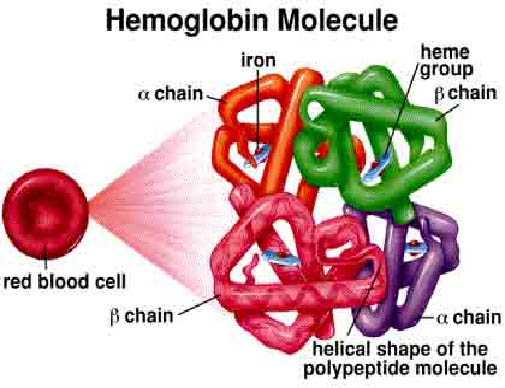
Image result for haemoglobin
Haemoglobin molecule


     WHAT IS HAEM??

v  Haem is actually an iron containing compound of porphyrin class which forms the non – protein of haemoglobin and some other molecule.

v  Haem synthesis occur largely in the mitochondria.
 
HOW ABOUT GLOBIN??

v  Superfamily of haem containing globular protein and involve in binding, transporting oxygen.

v  Globin synthesis occurs in the polyribosome.
 
WHAT IS HAEMOGLOBIN?

v   Haemoglobin is a protein molecule in the red blood cell that carries oxygen from lungs to the tissues and return carbon dioxide from tissue to lungs to be exhaled out. Molecular weight of the haemoglobin are about 68000 and comprise one third of a red cella. Haemoglobin is formed from the combination of haem and globin.65% of the haemoglobin will be synthesised in the erythroblast and the remaining 35% at the reticulocyte stage. A normal person would have haemoglobin HB-A that consist of four polypeptide chains a2B2 each with its own haem group. 
 
HAEMOGLOBIN SYNTHESIS


Image result for haemoglobin synthesis pathway
Haemoglobin synthesis

v  Haem synthesis begins with the condensation of glycine and succinyl-CoA to form δ-aminolevulinic acid (ALA). ALA then leaves the mitochondria and form porphobilinogen through a series of reaction forms coproporphyrinogen. This molecule then returns to the mitochondria and produce protoporphyrin.Proto-porphyrin is then combined with iron to form haem. Haem then exits the mitochondria and combines with the globin molecule which is synthesized in the ribosome.


   HAEMOGLOBIN CATABOLISM
    RED BLOOD CELL DESTRUCTION

 
Image result for red blood cell degradation
 

          Red cell destruction will occur after the life span of the red blood cell end (120 days)The cell are removed by macrophages of the reticularendothelial system in the bone marrow, liver and also spleen. This in turn leads to red cell metabolism gradually decline as enzyme are degraded and not replaced, until the cell become iron viable (the reason is unknown).The healthy red blood cells are in biconcave shape and after 120 days, the red blood cells become spherical in shape (change in morphology) resulting the spleen and other associate organs cannot recognise them and found that the cells could bring hazard to the body. The spleen then ask macrophage to destroy the cell.(terminology)

 
v  The breakdown of the cells release these 3 components:
 
-       Iron for recirculation via plasma transferrin to marrow erythroblast.

-       Protoporphyrin which is broken down to bilirubin.

-       Globin which are converted to amino acids.
 

     The bilirubin  circulates to the liver where it is conjugated to glucuronides which are excreted into the gut via bile and converted to the stercobilinogen and stercobilin (excreted in faeces). Stercobilinogen and stercobilin are reabsorbed back and excreted in urine as urobilinogen and urobilin. Meanwhile a small fraction of protoporphyrin is converted to carbon monoxide and excreted via the lungs. Globin chains are broken down to amino acid which are reutilized for general protein synthesis in the body.


Friday 24 March 2017

Erythropoiesis, Leukopoiesis & Thrombopoiesis

Image result for haemopoiesis
Pathway - Production of Rbc, Wbc and Platelets from the stem cells.


Erythropoiesis

  • Erythropoiesis is the process by which human erythrocytes are produced. It is triggered by erythropoietin, a kidney hormone produced during hypoxia.
  • Erythropoiesis takes place in the bone marrow, where hemopoietic stem cells differentiate and form proerythtoblasts and eventually shed their nuclei to become reticulocytes. Iron, vitamin B12, and folic acid are required for hemoglobin synthesis and normal RBC maturation.
  • Reticulocytes mature into normal, functional RBCs after 24 hours in the bloodstream.


Leukopoiesis

  • Process of making leukocytes, stimulated by various colony‐stimulating factors (CSFs), which are hormones produced by mature white blood cells. 
  • The development of each kind of white blood cell begins with the division of the hemopoietic stem cells into blast cells :
    1. Myeoblast divide to form :
      • Neutronophilic → Neutrophil
      • Eusinophilic → Eusinophil
      • Basophilic → Basophils
    2. Monoblasts lead to the development of monocyte
    3. Lymphoblast lead to the development of lymphocyte

Thrombopoiesis

  • Thrombopoiesis, the process of making platelets, begins with the formation of megakaryoblasts.
  • The megakaryoblasts divide without cytokinesis to become megakaryocytes, huge cells with a large, multilobed nucleus. 
  • The megakaryocytes then fragment into segments as the plasma membrane infolds into the cytoplasm.


Haemopoiesis

Image result for haemopoiesis cartoons

What is Haemopoiesis?

Haemopoiesis, or hematopoiesis, is the process by which new blood cells are formed. 

Bone marrow, the tissue inside bones, is one of the most active organs in the body, and is the site where red blood cells, the majority of white blood cells, and platelets are produced.

Site of haemopoiesis :
  • Fetus - in the yolk sac, liver or spleen and bone marrow according to the growth of pregnancy.
  • Children - the red marrow inside all of the bones makes blood cells.
  • Adults - vertebrae, skull, sternum, pelvis.
👷👷👷👷👷👷👷👷👷👷👷👷👷👷👷👷👷👷👷👷👷👷👷👷👷👷👷👷👷👷👷👷👷👷👷👷👷👷
(all the cells is produced in bone marrow in adult)

Extramedullary Haemopoiesis is production of blood cells outside of bone marrow
*It only happens in fetus stage.
*Can happen to adults if bone marrow is no longer functional or cannot keep up with the production
*Cause spleen and liver to enlarged.


Inside the bone marrow, cells called hematopoietic stem cells are able to produce all the different types of blood cells. 

They form either lymphoid stem cells or myeloid stem cells.

  • Lymphoid stem cells migrate to the spleen, lymph nodes and thymus and go on to produce lymphocytes, which are white blood cells involved in the immune system's response to infection. 
  • Myeloid stem cells develop into red blood cells, which carry oxygen, and white blood cells known as granulocytes, megakaryocytes and monocytes. Granulocytes and monocytes help fight off infection, while megakaryocytes break into fragments to form platelets, which are involved in blood clotting.


DEEP VEIN THROMBOSIS

DEEP VEIN THROMBOSIS .  DEFINITION     : Deep vein thrombosis occurs when blood clot forms and blocks a deep vein . Most commonly a ve...