Monday 27 March 2017

Haemoglobin synthesis and its catabolism

HAEMOGLOBIN SYNTHESIS AND ITS CATABOLISM

Image result for haemoglobin
Haemoglobin molecule


     WHAT IS HAEM??

v  Haem is actually an iron containing compound of porphyrin class which forms the non – protein of haemoglobin and some other molecule.

v  Haem synthesis occur largely in the mitochondria.
 
HOW ABOUT GLOBIN??

v  Superfamily of haem containing globular protein and involve in binding, transporting oxygen.

v  Globin synthesis occurs in the polyribosome.
 
WHAT IS HAEMOGLOBIN?

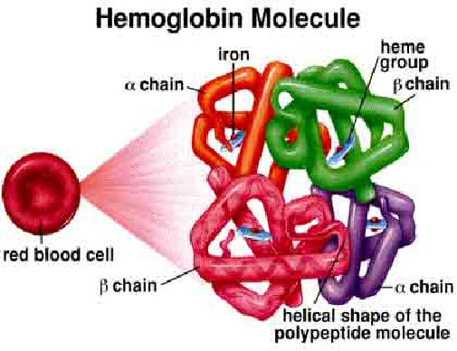
v   Haemoglobin is a protein molecule in the red blood cell that carries oxygen from lungs to the tissues and return carbon dioxide from tissue to lungs to be exhaled out. Molecular weight of the haemoglobin are about 68000 and comprise one third of a red cella. Haemoglobin is formed from the combination of haem and globin.65% of the haemoglobin will be synthesised in the erythroblast and the remaining 35% at the reticulocyte stage. A normal person would have haemoglobin HB-A that consist of four polypeptide chains a2B2 each with its own haem group. 
 
HAEMOGLOBIN SYNTHESIS


Image result for haemoglobin synthesis pathway
Haemoglobin synthesis

v  Haem synthesis begins with the condensation of glycine and succinyl-CoA to form δ-aminolevulinic acid (ALA). ALA then leaves the mitochondria and form porphobilinogen through a series of reaction forms coproporphyrinogen. This molecule then returns to the mitochondria and produce protoporphyrin.Proto-porphyrin is then combined with iron to form haem. Haem then exits the mitochondria and combines with the globin molecule which is synthesized in the ribosome.


   HAEMOGLOBIN CATABOLISM
    RED BLOOD CELL DESTRUCTION

 
Image result for red blood cell degradation
 

          Red cell destruction will occur after the life span of the red blood cell end (120 days)The cell are removed by macrophages of the reticularendothelial system in the bone marrow, liver and also spleen. This in turn leads to red cell metabolism gradually decline as enzyme are degraded and not replaced, until the cell become iron viable (the reason is unknown).The healthy red blood cells are in biconcave shape and after 120 days, the red blood cells become spherical in shape (change in morphology) resulting the spleen and other associate organs cannot recognise them and found that the cells could bring hazard to the body. The spleen then ask macrophage to destroy the cell.(terminology)

 
v  The breakdown of the cells release these 3 components:
 
-       Iron for recirculation via plasma transferrin to marrow erythroblast.

-       Protoporphyrin which is broken down to bilirubin.

-       Globin which are converted to amino acids.
 

     The bilirubin  circulates to the liver where it is conjugated to glucuronides which are excreted into the gut via bile and converted to the stercobilinogen and stercobilin (excreted in faeces). Stercobilinogen and stercobilin are reabsorbed back and excreted in urine as urobilinogen and urobilin. Meanwhile a small fraction of protoporphyrin is converted to carbon monoxide and excreted via the lungs. Globin chains are broken down to amino acid which are reutilized for general protein synthesis in the body.


Friday 24 March 2017

Erythropoiesis, Leukopoiesis & Thrombopoiesis

Image result for haemopoiesis
Pathway - Production of Rbc, Wbc and Platelets from the stem cells.


Erythropoiesis

  • Erythropoiesis is the process by which human erythrocytes are produced. It is triggered by erythropoietin, a kidney hormone produced during hypoxia.
  • Erythropoiesis takes place in the bone marrow, where hemopoietic stem cells differentiate and form proerythtoblasts and eventually shed their nuclei to become reticulocytes. Iron, vitamin B12, and folic acid are required for hemoglobin synthesis and normal RBC maturation.
  • Reticulocytes mature into normal, functional RBCs after 24 hours in the bloodstream.


Leukopoiesis

  • Process of making leukocytes, stimulated by various colony‐stimulating factors (CSFs), which are hormones produced by mature white blood cells. 
  • The development of each kind of white blood cell begins with the division of the hemopoietic stem cells into blast cells :
    1. Myeoblast divide to form :
      • Neutronophilic → Neutrophil
      • Eusinophilic → Eusinophil
      • Basophilic → Basophils
    2. Monoblasts lead to the development of monocyte
    3. Lymphoblast lead to the development of lymphocyte

Thrombopoiesis

  • Thrombopoiesis, the process of making platelets, begins with the formation of megakaryoblasts.
  • The megakaryoblasts divide without cytokinesis to become megakaryocytes, huge cells with a large, multilobed nucleus. 
  • The megakaryocytes then fragment into segments as the plasma membrane infolds into the cytoplasm.


Haemopoiesis

Image result for haemopoiesis cartoons

What is Haemopoiesis?

Haemopoiesis, or hematopoiesis, is the process by which new blood cells are formed. 

Bone marrow, the tissue inside bones, is one of the most active organs in the body, and is the site where red blood cells, the majority of white blood cells, and platelets are produced.

Site of haemopoiesis :
  • Fetus - in the yolk sac, liver or spleen and bone marrow according to the growth of pregnancy.
  • Children - the red marrow inside all of the bones makes blood cells.
  • Adults - vertebrae, skull, sternum, pelvis.
👷👷👷👷👷👷👷👷👷👷👷👷👷👷👷👷👷👷👷👷👷👷👷👷👷👷👷👷👷👷👷👷👷👷👷👷👷👷
(all the cells is produced in bone marrow in adult)

Extramedullary Haemopoiesis is production of blood cells outside of bone marrow
*It only happens in fetus stage.
*Can happen to adults if bone marrow is no longer functional or cannot keep up with the production
*Cause spleen and liver to enlarged.


Inside the bone marrow, cells called hematopoietic stem cells are able to produce all the different types of blood cells. 

They form either lymphoid stem cells or myeloid stem cells.

  • Lymphoid stem cells migrate to the spleen, lymph nodes and thymus and go on to produce lymphocytes, which are white blood cells involved in the immune system's response to infection. 
  • Myeloid stem cells develop into red blood cells, which carry oxygen, and white blood cells known as granulocytes, megakaryocytes and monocytes. Granulocytes and monocytes help fight off infection, while megakaryocytes break into fragments to form platelets, which are involved in blood clotting.


DEEP VEIN THROMBOSIS

DEEP VEIN THROMBOSIS .  DEFINITION     : Deep vein thrombosis occurs when blood clot forms and blocks a deep vein . Most commonly a ve...